Abducens Palsy


|
Abducens Palsy |
|
In the first example, an abducens palsy of the right eye is simulated. The abducens palsy is an incomitant form of strabismus, i.e. the squint angle increases in the main functional direction (towards abduction in this pathology) of the affected muscle - the lateral rectus muscle. Clinically, the patient increasingly shows double images (uncrossed) towards abduction, thus when looking to the right. Possible causes for an abducens palsy are, among other things, traumata of the peripheral nerves (that is the entire nerve without the nucleus) caused for example by a basal skull fracture. As a result, a damage to the nerve somewhere from the nucleus (in the brainstem) up to the insertion can occur. In case of a damage directly within the area of the nucleus, it is possible that also the nearby interneurons (the connections between the nucleus of the nervus abducens and the conjugated muscle on the other side à medial rectus muscle) are hurt. This case is not assumed in this example.
Simulation of the Pathology
As a consequence of the damage to the nerve, the contractile strength of the muscle has to be reduced. This is the basis for the simulation.
First a new patient is created. To do so, the menu item "Patient" - "New Patient" is used, which displays the following dialog:
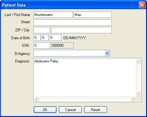
In this dialog, information about the patient can be entered. After clicking on the "OK" button, the new patient is created and the program is ready for starting with the simulation.
In order to successfully simulate the abducens palsy, the contractile strength of the lateral rectus muscle of the right eye has to be reduced. In SEE++, this force reduction is carried out in the muscle data dialog, which is accessible either by the menu item "Data" - "Right Eye" - "Muscles" or by the tree in the left part of the program window by using the tree item "Muscle Data" in the section for the right eye. In the upper area of the appearing window there are several "tab sheets", namely one containing general parameters and one for each muscle. By clicking on the tab sheet "Lateral Rectus", the dialog now looks like this:
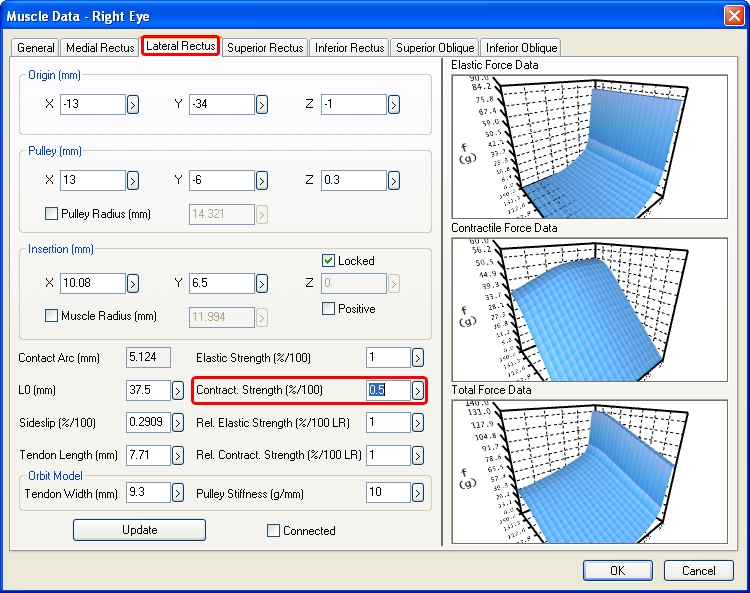
In the field "Contract. Strength" the strength of the muscle can be reduced by changing the value from 1 %/100 to 0.5 %/100 and afterwards closing the dialog by clicking on "OK".
When the Hess-diagram is recalculated now, the modifications from the muscle data dialog are immediately taken into account. By default, the Hess-diagram for the left eye (right eye fixing) is displayed in the diagram-window with the number 1. After the calculation of the Hess-diagram has been successfully finished, the following is displayed:
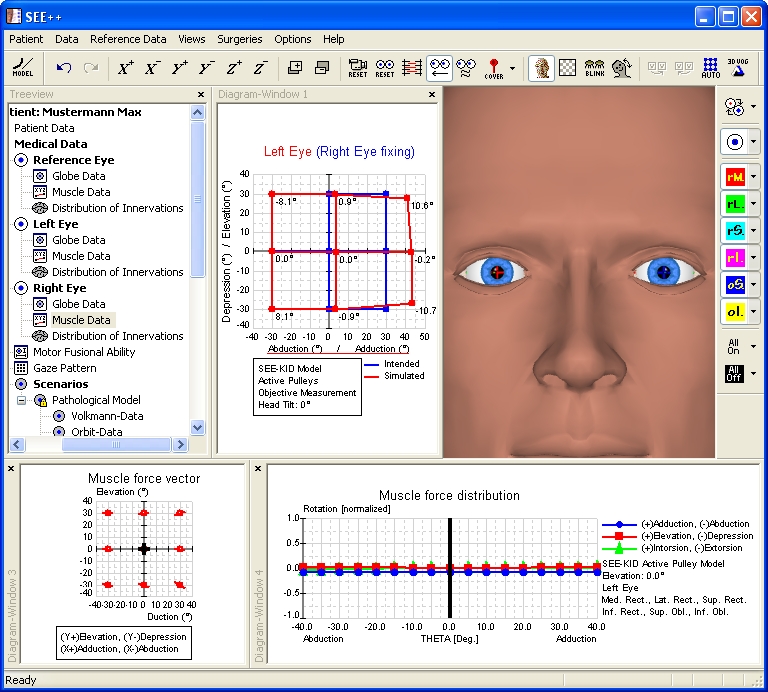
In the calculated Hess-diagram the exceeding reaction of the following left eye (right eye fixing) into adduction can be clearly seen. In order to display the second Hess-diagram for the right eye (left eye fixing), use the menu item "Views" - "Diagram-Window 2". By default, the now displayed diagram-window with the number 2 displays the Hess-diagram for the right eye (left eye fixing). If this is not the case, you can simply click into one of the diagram-windows with the right mouse button and select the desired diagram in the displayed popup menu by left-clicking on it. If the size of the two diagram-windows is adjusted accordingly by clicking on the dividing line between the diagrams and resizing the diagrams with the left mouse button pressed, the following view is displayed:
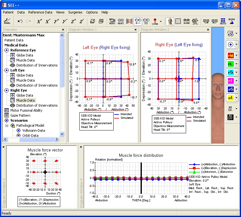
In the Hess-diagram for the right eye (left eye fixing) the restriction of the right eye in abduction can be clearly seen. On the basis of the results of the simulation, which are shown in the Hess-diagrams, the simulation of the abducens palsy can be considered as finished.
Surgical Correction
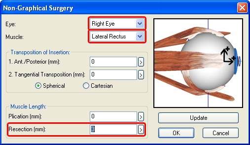
For the surgical correction of the simulated abducens palsy, a strengthening of the paretic muscle is necessary. The surgery is carried out by shortening (resection) the affected lateral rectus muscle. During a resection surgery, the insertion of a muscle is separated from the globe, a piece of the muscle is cut off and afterwards, the muscle is fixed again at the same position on the globe. For shortening a muscle in SEE++, the "Non-Graphical Surgery" dialog is used, which is accessible by the menu item "Surgeries" - "Non-Graphical Surgery". After the dialog has been opened, the desired eye and the desired muscle are selected, in this case the right eye and the lateral rectus muscle. Now a resection of 3 mm of the selected muscle is carried out by entering the appropriate value in the field "Resection (mm)". The following picture shows the dialog with the entered resection value:
After closing the dialog with a click on the "OK" button, the program recalculates the two Hess-diagrams. When the calculation is finished, the following picture is displayed:
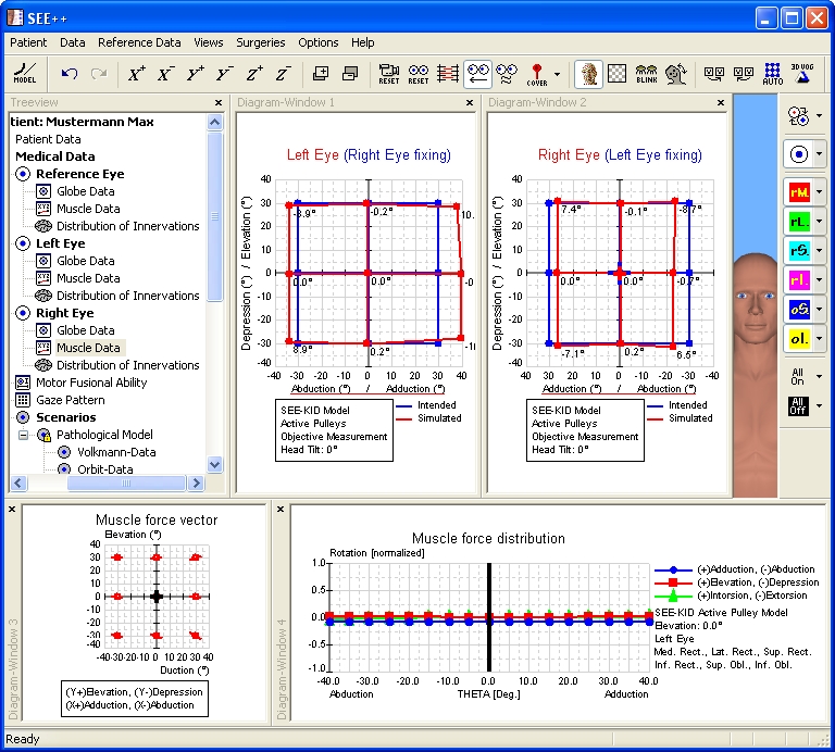
Now the two Hess-diagrams show that the goal of the surgical correction has been achieved, namely to get the double image free zone into the primary position without substantially weakening the adduction. However, a complete "healing" of the palsy by modifying the innervational component in the model is clinically not possible, since a surgical modification of the innervation is impossible.