Simulation


|
Simulation |
|
SEE++ uses a biomechanical model for simulating the binocular Hess-Lancaster test. The results of this test can be visualized in form of Hess-diagrams (left eye fixing and right eye fixing) and in form of a textual view (squint-angles diagram).
The main difference between the two visualizations is the declaration of the deviations. While the Hess-diagram offers a visual impression of a pathological situation and can be calculated for freely chosen gaze positions, the squint-angles diagram offers a textual illustration of the deviation values in the nine main gaze directions.
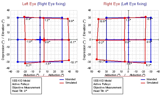
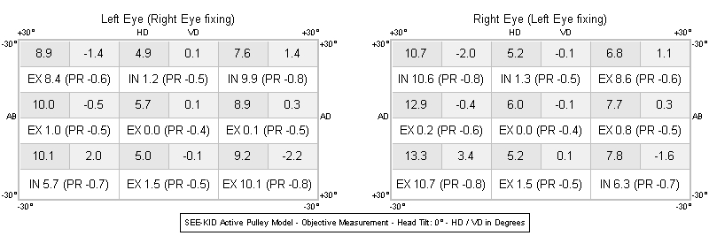
Hess-Diagrams and Squint-Angles Diagram (Right Eye Fixing and Left Eye Fixing)
In the squint-angles diagram the particular deviations for each gaze direction for right eye fixing and left eye fixing are entered, whereby HD denotes horizontal deviation and VD denotes vertical deviation. In the white fields of the diagram the torsional deviation as excyclotorsion or incyclotorsion (EX or IN) and the protrusion in mm (PR; negative protrusion = retraction) are specified. All values apart from the protrusion are in degrees. For HD and VD the user can select whether the values should be specified in degrees or prism diopters (PD).
The signs for the horizontal deviations are defined as follows:
•+HD: Towards the nose (adduction)
•-HD: Away from the nose (abduction)
The signs for the vertical deviations are defined differently for right eye and left eye fixing:
Right Eye Fixing
•+VD: Downward deviation of the left eye (depression)
•-VD: Upward deviation of the left eye (elevation)
Left Eye Fixing
•+VD: Upward deviation of the right eye (elevation)
•-VD: Downward deviation of the right eye (depression)
Different Types of Torsion (Cyclorotation)
The torsion values shown in the Hess-diagrams and the squint-angles diagram of SEE++ depend on the type of torsion measurement chosen in the program. By default, the torsion values in the diagrams correspond to clinical patient data resulting from an objective measurement. In this case objective measurement refers to a measurement, where the eye position including the torsion is directly measured on the patient (e.g. with a VOG (video oculography) system or with search coils) without the need for any information provided by the patient. If one now switches to subjective torsion measurement in SEE++, then the torsion values correspond to measurement values stated by the patient during a subjective test (e.g. tangent screen, Harms' wall). These torsion values correspond to the difference the patient perceives between one and the other eye (cyclo-deviation), since the "normal" torsion according to Listing's law (e.g. 8.2 degrees when looking 30 degrees in adduction and 30 degrees in elevation) also exists in a healthy eye (see Listing's law) and is not subjectively perceived by the patient. In order to be able to compare clinically measured torsion values with simulated torsion values, the appropriate torsion measurement has to be selected in the program.
In addition to Listing's torsion, SEE++ also calculates the counter roll, which occurs during the head tilt towards the left or right shoulder (VOR - vestibulo ocular reflex). For this purpose, the appropriate counter roll is calculated based on the current head tilt of the "virtual" patient and is combined with Listing's torsion or the current cyclo-deviation (depending on the selected torsion measurement). The combined torsion is then displayed in the different diagrams. Moreover, the Stateviewer offers the possibility to examine the separate components of which the combined torsion shown in the diagrams consists.
Simulation Task Flow
Sometimes it may be helpful to know how the internal simulation task flow of SEE++ works in order to interpret a simulation result. The following figure shows the schematic simulation task flow for the Hess-Lancaster test, which SEE++ performs for each gaze position of the fixing eye gaze pattern.
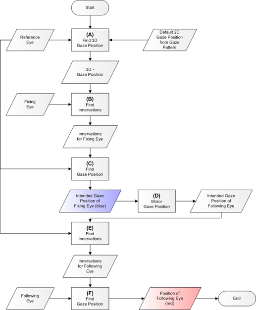
Simulation Task Flow for the Hess-Lancaster Test (Based on Orbit™, See [Miller, 1999])
The goal is to calculate the deviations of the following eye from the fixation positions of the fixing eye. Thereby, the fixing and following eye can be exchanged, i.e. either the right eye is fixing and the left eye is following or the left eye is fixing and the right eye is following. This makes it possible to calculate the Hess-diagram for right eye fixing and left eye fixing.
| (A) | Starting from a healthy reference eye and a given fixation position, a complete 3D eye position is calculated. |
| When providing a fixation position, of course only abduction/adduction and elevation/depression can be specified. The specification of a torsion is not possible, since a patient cannot explicitly rotate the eye around a specified torsional angle. As a result, the complete position of the fixing eye has to be calculated by generating innervations for eye positions with Listing torsions with the help of the reference eye until the position of the fixing eye matches with these innervations and the desired 2D eye position. |
| (B) | The innervation pattern for all six eye muscles is calculated for the determined 3D eye position with the fixing eye, which is necessary to bring the fixing eye into the desired position. |
| (C) | These innervations are now put into the reference eye in order to calculate its eye position. If the fixing eye is pathological, the eye position calculated with the reference eye differs from the previously calculated eye position. The result of this calculation is now the actually intended fixation position of the fixing eye. |
| (D) | In this step, the intended fixation position of the fixing eye is mirrored in order to get the intended position of the following eye. Since innervations cannot be mirrored (the contralateral synergist of the right lateral rectus muscle is the left medial rectus muscle which may not act in exactly the same way), this "indirection" of mirroring the eye position is necessary. The mirroring of an eye position is possible, because abduction/adduction and torsion of the two eyes have different signs for describing the same direction. |
| (E) | Now the innervations of the reference eye are determined from the mirrored eye position so that the position of the following eye can be calculated afterwards. |
| (F) | The innervations of the reference eye are now used to calculate the position of the following eye. This position is one of the red points in the Hess-diagram. |
Literature
[Brugger, 2000] P.C. Brugger. Der 3D Anatomie Atlas. Weltbild Verlag GmbH, Augsburg, Deutsche Erstausgabe, 2000.
[Buchberger and Mayr, 2000] M. Buchberger and H. Mayr. SEE-KID: Software Engineering Environment for Knowledge-based Interactive Eye Motility Diagnostics. In Proceedings of International Symposium on Telemedicine, Gothenburg, Sweden, 2000.
[Clark et. al., 2000] R.A. Clark, Miller J.M. and J.L. Demer. Three-dimensional location of human rectus pulleys by path inflections in secondary gaze positions. Investigative Ophthalmology & Visual Science, 41(12): 3787–3797, November 2000.
[Demer et. al., 2000] J.L. Demer, S.Y. Oh and V. Poukens. Evidence for Active Control of Rectus Extraocular Muscle Pulleys. Investigative Ophthalmology & Visual Science, 41(6):1280–1290, May 2000.
[Günther, 1986] S. Günther. Die modellmäßige Beschreibung der Augenmuskelwirkung. Diploma Thesis, University Hamburg, Universitätskrankenhaus-Eppendorf, Abteilung für medizinische Optik, 1986.
[Kaufmann, 1995] H. Kaufmann. Strabismus. Ferdinand Enke Verlag, Stuttgart, 2. Edition, 1995.
[Miller 1999] J.M. Miller. Orbit™ 1.8 Gaze Mechanics Simulation Users Manual. Eidactics, Suite 404, 1450 Greenwich Street, San Francisco, CA 94109, USA.
[Miller and Demer, 1996] J.M. Miller and J.L. Demer. Uses of Biomechanical Modeling. In Proceedings of CLADE, Buenos Aires, 1996.
[Miller and Demer, 1999] J.M. Miller and J.L. Demer. Clinical Applications of Computer Models for Strabismus. In eds Rosenbaum, A and Santiago, AP, Clinical Strabismus Management. Philadelphia, W. B. Saunders.
[Pschyrembel, 1994] Pschyrembel. Klinisches Wörterbuch. Nikol VerlagsgmbH, Hamburg, 257. Edition, 1994.
[Schäffler and Schmidt, 1998] A. Schäffler and S. Schmidt. Biologie, Anatomie und Physiologie. Urban und Fischer Verlag, Munich, 3. Extended Edition, 1998.
[Yanoff and Duker, 2003] M. Yanoff and J.S. Duker. Ophtalmology. Mosby, London, 2. Edition, 2003.